The advent of new technologies has led to a surge in publications in the field of single cell and spatial transcriptomics, including relating to neurodegeneration. This has allowed researchers to conduct integration analysis. Isar Nassiri of the Parkinson’s Neuropathology Group, University of Oxford, outlines in this presentation a study exploring the neuronal vulnerability of neurons in Parkinson’s disease using the integration of spatial and single cell data.
Single cell and single nuclei approaches have the advantage of distinguishing between different cell types in their gene expression profile. Nassiri’s team prefer to use single nuclei (sn)RNA-seq over single cell (sc)RNA-seq due to the nuclei’s better resilience as compared to the whole cell. Further advantages include more integrated RNA, especially for older samples, and a reduced cell size bias. scRNA-seq can also run into dissociation issues with older samples, which snRNA-seq avoids.
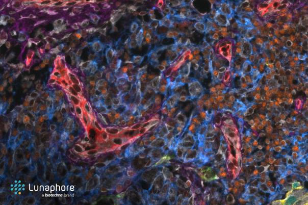
Nassiri then outlined the various spatial transcriptomic techniques available for mapping cells and studying their associated pathology. This provides a spatial context for gene expression, overlaying the single cell data on top of the tissue section. In situ hybridisation uses a fluorescent label and microscopy to detect gene expression, in situ sequencing uses a padlock probe and ligation to do this, in situ capture uses a system of spots containing reverse transcript primer probes for gene expression detection.
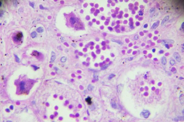
The experimental plan that Nassiri’s team went with used a control and Parkinson’s samples from the Oxford Biobank. Then, they performed sectioning and staining using methods like immunofluorescence and H&E to reveal the pathological hallmarks. In parallel, the team conducted spatial barcoding and sequencing, providing the metrics for expression activity which can then be overlayed on top of the tissue section.
The results revealed significant degradation in the substantia nigra region of the brain and highlighted the molecular mechanisms behind neuron vulnerability. By integrating spatial and single-cell data, the study was able to annotate neurons with and without Lewy body pathology. This allowed for a detailed comparison of their expression patterns and provided insights into the molecular mechanisms that make certain neurons more vulnerable to Parkinson's disease.