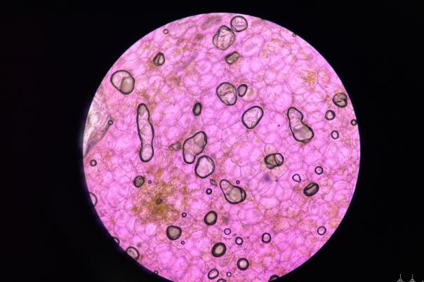
The central question for our AI in Spatial Biology interest group webinar, which took place in June 2026, was: how can AI help spatial biology move from complex tissue images to trusted, interpretable, and clinically relevant insight?
The webinar was facilitated by Tommy Ming Tang, Director of Bioinformatics at AstraZeneca. Ming brought a translational and computational perspective on how spatial and pathology data can support discovery, biomarker research, and clinical development.
The session was also facilitated by Egon J. Ranghini, Senior Science Technology Advisor at 10x Genomics. Egon brought deep expertise in spatial biology platforms, data generation, and the technologies helping researchers move from complex tissue images to meaningful biological data.
The meeting brought together a broad cross-section of the spatial biology, computational pathology, translational science, and biomarker communities. Participants included senior scientists, bioinformaticians, pathologists, biomarker leads, data scientists, clinical research as well as business development leaders. Organisations represented included pharmaceutical and biotechnology companies, academic research institutes, hospitals, technology providers, and specialist bioinformatics and data organisations.
This diversity of attendees reflected the interdisciplinary nature of the challenge: AI in spatial biology is not only a computational issue, but also a biological, clinical, technical, operational, and translational one.
Ahead of the meeting, participants were surveyed on how they currently use AI, where they face the greatest challenges, and what would accelerate adoption.
The results showed a clear problem: while AI is already being used across parts of the spatial biology pipeline, confidence in AI-driven outputs remains limited. Respondents reported using AI for image segmentation and cell detection, feature extraction, cell type annotation, downstream analysis, and multi-modal data integration. However, some respondents said they are not yet using AI, showing that adoption remains uneven.
The biggest bottleneck identified was biological interpretation — the challenge of moving from image-derived data to meaningful biological or clinical insight. Other pain points included segmentation accuracy and data integration across platforms.
Confidence was also cautious. No respondent reported high confidence in AI-driven outputs. Most were either somewhat confident or had low confidence, highlighting the need for better validation, reproducibility, and quality control.
When asked about barriers to clinical adoption, respondents pointed to data standardisation, integration with pathology workflows, and lack of validation or reproducibility. The biggest technical challenges were computational cost and infrastructure, lack of annotated data, model generalisation across datasets, and data normalisation. The most common request was for more high-quality training data, followed by clearer best practices and cross-platform standards.
These survey findings framed the purpose of the meeting: to explore not only where AI can add value, but how the field can build confidence in AI-driven outputs through stronger data quality, validation, standardisation, and biological interpretation.
The webinar explored how artificial intelligence is being applied across the spatial biology pipeline, from image analysis and cell detection through to data interpretation, clinical development, and precision medicine.
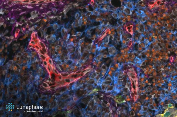
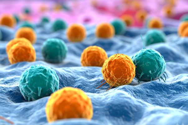
A key theme was that AI is already proving useful in the early stages of spatial biology analysis. Egon highlighted its role in cell segmentation, cell annotation, and multi-modal data integration, particularly where researchers need to move from complex tissue images to structured, usable data. He noted that segmentation remains especially challenging in irregular cell types and complex tissues, where AI models need high-quality training data to improve accuracy.
Ming emphasised that the real challenge is not only generating spatial data, but making it reliable, interpretable, and useful for clinical or translational applications. He pointed to the cost of spatial technologies, the difficulty of standardising workflows, and the need to validate AI outputs against real biological and clinical outcomes.
Several participants reinforced the importance of data quality. One attendee raised the need for large, high-quality datasets and questioned how AI could be used beyond segmentation, including for quality control and detecting technical artefacts. Another participant highlighted the importance of standardised data pre-processing, warning that spatial datasets can vary significantly depending on platform, workflow, and analysis methods.
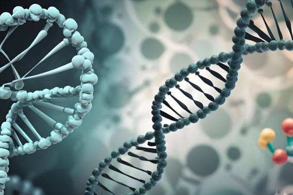
The meeting also moved beyond technology into biological interpretation. One contributor shared how AI and large-scale transcriptomic datasets are being used to identify disease mechanisms and support targeted therapy development. They stressed that AI must be grounded in biological understanding, precision, and rigorous validation, particularly when applied to medicine.
Overall, the meeting made clear that AI has significant potential to accelerate spatial biology and computational pathology. However, the field must address several practical barriers: data quality, annotation, computational cost, platform standardisation, biological interpretation, and clinical validation.
The central message was that AI should not be viewed as a black box or a shortcut. Its value depends on high-quality data, clear biological questions, robust validation, and close collaboration between computational scientists, biologists, pathologists, and clinical teams.
As spatial biology moves from research into translational and clinical settings, the challenge will be to ensure that AI-generated outputs are not only accurate, but also meaningful, reproducible, and actionable.