Key Takeaways
- SMMILe accurately maps and quantifies tumour regions and aggressiveness on pathology images.
- SMMILe outperforms existing AI tools using only simple patient-level labels for training.
- The tool could offer more personalised cancer treatment by revealing detailed tissue architecture.
Introduction
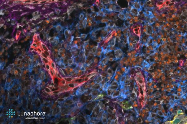
Researchers University of Cambridge have developed a machine learning algorithm that facilitates accurate spatial quantification of tumour tissue on digital pathology images. This major breakthrough could enable personalised treatment decisions guided by what the tumour looks like and what biological insights it reveals.
SMMILe (Superpatch-based Measurable Multiple Instance Learning) does not only exceed the performance of current state of the art whole slide image (WSI) tissue classification tools for the detection of cancer cells in tumour biopsies but also predicts where tumour lesions are located and the proportion of regions with different levels of aggressiveness.
Instead of classifying a slide, SMMILe goes a step further by measuring how different tumour subtypes, grades and surrounding tissue components are organised which gives a structures and truly quantitative view of the tissue.
Methodology
Zeyu Gao, the lead researcher on the project and his team trained SMMILe using slides that had been given simple, patient-level diagnostic labels, such as cancer type or grade rather than needing time-consuming, detailed region-by-region annotations from pathologists. The researchers tested the algorithm on eight datasets comprising 3850 whole-slide images covering lung, kidney, ovarian, breast, stomach, and prostate cancer.
Analysis
Next, they compared SMMILe’s performance with nine other WSI classification analysis AI tools. The analyses showed that SMMILe’s performance in metastasis detection, subtyping, and grading either matched or exceeded these tools at slide-level classification, while significantly outperforming them when it came to estimating the proportions and spatial distribution of lesions.
Gao said: “SMMILe moves pathology from qualitative impressions to precise spatial quantification,” he continued, “Patients who look similar under conventional pathology can now be distinguished by their tissue architecture and spatial organisation. This provides a new layer of information to guide personalised therapies.”
References