Marie Laviron’s lab is interested in the resident macrophages of the liver called Kupffer cells. In particular, her team investigates the signals that drive the development, maintenance and function of these macrophage populations. These signals are defined together as the macrophage ‘niche’, which in the liver includes stellate cells, hepatocytes, and endothelial cells. The cells within the niche deliver signals to Kupffer cells which plays a major role in defining their identity.
In order to define these cells, the team first needs to understand the tissue they are working with. This is why her team, in a massive collaborative project, have generated an atlas of cells, gene and protein expression, and spatial distribution within the liver at a single cell level.
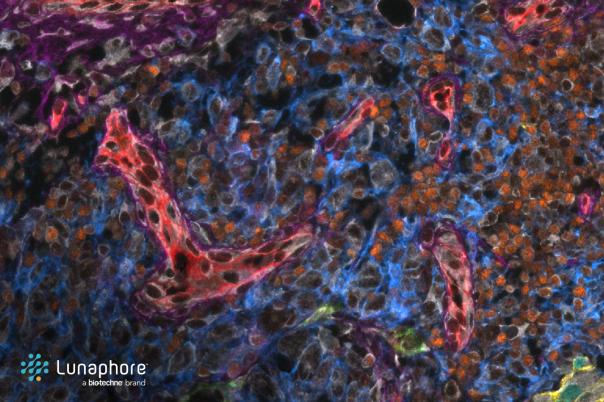
To create this atlas, the team performed CITEseq to get transcriptomic and protein signals for all the cells in the donor liver. They combined mRNA and protein data to achieve the broadest span of data that they could. From there, they were able to identify clusters, distinguish subsets, and identify markers within those subsets. Laviron’s team took those markers and used them with spatial imaging techniques to locate those populations.
Focusing on Kupffer cells, they pinpointed Cd5l and VISIG4 as highly conserved markers of Kupffer cells in many species. They found that using both protein and mRNA staining together was essential to identifying these populations.
To obtain deeper transcriptomic data, the researchers used Visium combined with a bioinformatic algorithm to cartograph zones of the liver related to their metabolic function. Depending on which zone you look at in the liver, you’ll find different populations of macrophages.
Using the mRNA and protein staining, the team identified spatial interactions between Kupffer and stellate cells in the niche. This was an interaction which they found was crucial for Kupffer cell identity.
From here, the researchers wanted to look at the other interactions that defined the identity of Kupffer cells. To do this, they used Miltenyi MACSima technology which is able to do 100 plex proteomics via a cycle of staining and bleaching. From there they saw Kupffer cells interacting with B cells, which the team are now investigating.
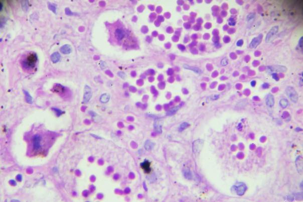
Putting all their data together generated a highly detailed map of the liver biology. This was translated into human samples which showed conservation of the structures and markers from their mouse samples.
The second half of Laviron’s presentation detailed how her lab used the atlas to investigate what happens to the Kupffer cell niche upon liver metastasis. It revealed that Kupffer cells are excluded from the tumor nodule and replaced by monocyte-derived tumor-associated macrophages.