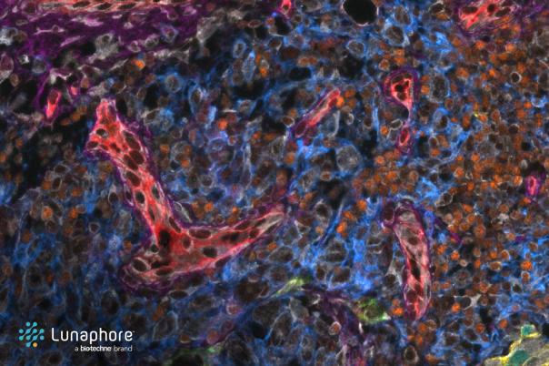
Min Xu, Co-Director MS in Computational Biology Program, PI & Assistant Professor, Carnegie Mellon University, highlighted his recent work on extracting structural and spatial organisation information of macromolecules inside the single cells captured by cryo-electron tomography. When zooming into a sub-region of the cell, one can observe a lot of macromolecules. The different structural and spatial organisations of these macromolecules influence their interactions with other sub-cellular structures. Xu’s method involves freezing cell samples and using electron beams to obtain projection images from different angles.
Originally lack of data acquisition methods meant it was hard to capture such behaviours, but recent advancements of cryo-electron tomography have made the acquisition of this method possible and promising. Xu gave an example of a 3D representation of these cells and showed that with sub-molecular resolution scientists can obtain insight into macromolecular structure.
The segmentation and identification processes are typically laborious, and when done manually, structural biologists and quality imaging experts can spend weeks or months processing a single chromatogram. Therefore, Xu emphasised the importance of automating these tasks to improve efficiency and accuracy and free up scientists’ time.
A tomogram is a 2D image built up from a series of virtual slices through the body. In practice, Xu and his team may have to process millions of subtomograms, and existing methods are too slow to process such large amounts of data. To handle the large volume of data and improve the speed and accuracy of subtomogram classification, Xu has invested in structural pattern mining. He has developed deep learning-based methods that leverage convolutional neural networks to classify subregions of tomograms without relying on external templates.
Xu outlined the outcome of using this method: “We are able to achieve a significant improvement of both speed and accuracy compared to the previous large high throughput subtomogram classification methods that are geometrically based.”
Several challenges, including high levels of noise, missing data, and the crowded nature of cytoplasm in tomograms, still remain. To improve his method, Xu is investigating design visualisation methods for debugging processes. On top of this, the team is seeking to reduce the number of annotations to classify and identify specific structures and improve the robustness of deep clustering methods.