Multiplexed imaging is frequently used in translational research, particularly when studying cancer. Jia-Ren Lin, Technical Director of the Tissue Imaging Platform at Harvard Medical School, aimed to investigate the applicability of multiplexed imaging in understanding cell-cell interactions and spatial organisation of cells within tumours. However, Lin also mentioned that he wants to go a step further and apply multiplexed imaging to other diseases.
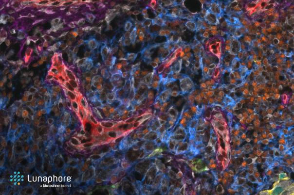
Lin introduced cyclic immunofluorescence as one of the most fundamental imaging approaches in antibody-based spatial proteomics. Cyclic immunofluorescence is a straightforward protocol involving iterative staining and imaging of tissues.
Antibody validation is a crucial process but highly challenging, especially in tumour tissue. Furthermore, there are often discrepancies when translating findings from model systems to human tissues. To combat this, Lin tried using principles such as tissue specificity, localisation, and sample cell specificity to guide his investigation. He suggested using an antibody on top of an antibody procedure. In other words, he advocated for using well-validated antibodies to cross-validate other antibodies. He proposed using CD3 to cross-validate CD8.
PD-L1, a central marker in immuno-oncology. Lin discussed the discrepancies between different PD-L1 antibodies used for diagnosis, and efforts to understand these differences using mass spectrometry and spatial proteomics.
A case study used serial sections of colorectal cancer tissue to reconstruct 3D tissue structure and produce this in a single view. In the 3D view, they found an isolated PLS structure that appeared finger-like. Whereas in 2D, it appeared as an isolated ball. This illustrated the compositional differences across tumour regions. Furthermore, the findings suggest that PD-L1 is often expressed in immune cells near tumours like macrophages and dendritic cells rather than in tumour cells themselves, challenging conventional assumptions.
From a clinical standpoint, Lin revealed the Orion platform, a multiplex imaging system capable of aligning high-resolution histology with immunofluorescence data. This platform was used to replicate and enhance the ImmunoScore, a prognostic tool for colorectal cancer based on CD3 and CD8 expression. The team developed hundreds of new scoring combinations with potentially better prognostic power, validated in two independent cohorts.