Crohn’s disease is a chronic inflammatory disease affecting the gastrointestinal tract. The disease causes severe symptoms of pain and discomfort in the millions living with it worldwide. In this presentation, Priyank Patel, a Senior Scientist at Boehringer Ingelheim, focused on understanding Crohn’s disease using spatial proteomics.
Inflammation and resolution in Crohn’s are driven by myeloid cells from the very earliest stages of the disease. Bacterial dysbiosis can cause myeloid cells to initiate innate immune sensing mechanisms and inflammation due to immune reaction against imbalanced microbes. When the bacterial load is cleared, myeloid cells also drive resolution and epithelial barrier repair.
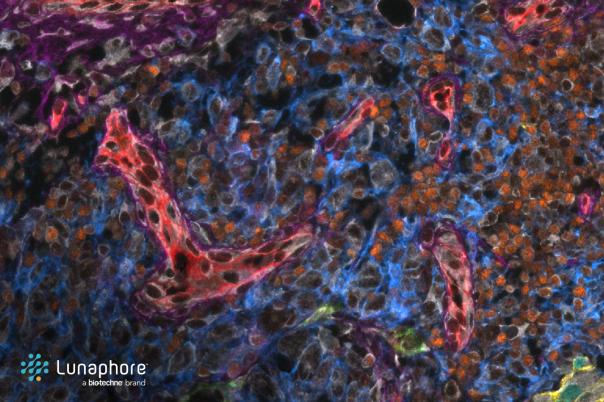
To understand the heterogeneity of these myeloid cells, Patel and his team have worked on optimising a 38 protein marker immunohistochemistry panel. The panel was developed using the Luna Force Comet platform, a traditional immunofluorescence platform based on cycles of staining, imaging, and elution in an automated workflow. They performed this staining on 11 tissues from four Crohn’s disease patients
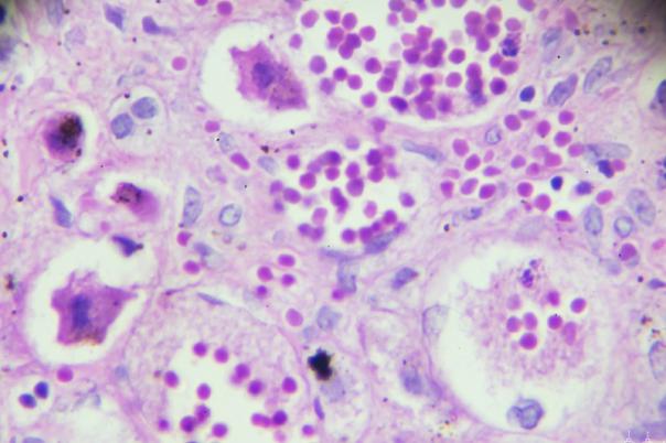
Before discussing the data, Patel noted that FFPE tissues are made up of diverse niches of cells with unique phenotypes. These niches are spread all across the tissue sample. Therefore, the team’s first task was to group together the niches that exhibit unique pathologies.
They identified 14 meta clusters, which were analysed to understand cellular heterogeneity and disease context. Analysis was divided into single cell analysis to identify cellular heterogeneity and pathology work to characterise the 14 meta clusters and study target expressions within these pathologies.
The expression patterns of two targets were studied across different clusters and cell populations, revealing distinct expression levels and cell types involved. Spatial analysis of macrophages expressing the targets showed different phenotypes and functions within various niches, indicating potential disease relevance.
Pathological insights helped define the clusters and their associated pathologies, providing context for target expressions and guiding further exploration. The next steps involve transferring findings to in vitro teams for phenotypic confirmation, automating pathological analysis, and integrating spatial transcriptomics and proteomics data for deeper insights.