The team at the Microscopy and Histology Core at La Jolla Institute for Immunology specialise in developing microscopy techniques and image analysis. The seven members of the group have a total of 96 years of combined experience, producing 15,000 slides annually – around 185lbs of glass. In this presentation, Zbigniew Mikulski, Director of the Core, walks us through the challenges that the group faces, and the solutions they have developed to tackle them.
Mikulski began the presentation by discussing the challenges that the team often face when spatial imaging. These issues include autofluorescence, cross-reactivity between antibodies, and limited numbers of channels in microscopes. He said that different technology providers can offer various solutions to these problems.
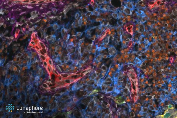
In spatial imaging, there are various factors that the team consider when deciding on a platform to use. Most important for Mikulski’s group is image quality, due to the fact that they would be performing chemical analysis later down the line. Mikulski said that the important aspects of image quality they look for are dynamic range, resolution, and minimal artifacts, with a focus on both bright and dim signals for accurate analysis.
Academic labs face budget constraints, making the cost of equipment, service contracts, staff time, and antibodies crucial factors in technology selection. As such, with limited resources, the group had to consider assay setup time, cost per slide, and compatibility with existing workflows when choosing their platforms.
The presentation then outlined the staining workflow. This involves baking slides, deparaffinisation, antigen retrieval, and a unique bleaching step to reduce autofluorescence without harming antigens. Designing and optimising panels involves testing primary antibodies, analysing signal intensity, and titrating conjugated antibodies, with a focus on cost-effectiveness and customization.
Proper fixation, understanding expected results, and the ability to experiment without high costs are key takeaways, with an emphasis on sharing successes and failures within the community.