The Cambridge Institute has 21 research groups, all dedicated to various aspects of cancer research and supported by 12 core facilities with high-end equipment and expert scientists. Cara Brodie, Senior Scientific Associate, University of Cambridge, works in the histopathology and ISH core facility, which is centred on routine histopathology, immunohistochemistry, and in situ hybridisation.
Within the institute, there has been a massive push for the implementation of spatial biology techniques. Brodie sought to uncover not just the genomic and protein data within the tissue but also how the spatial context influences cancer development.
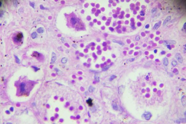
So, the team launched a project that aimed to identify glioblastoma subtypes and their metabolic properties using carbon-13 labelled glucose infusion. This was followed by mass spectrometry, H&E staining, and imaging mass cytometry (IMC). The IMC uses metal isotope tags instead of fluorophores, allowing for the identification of multiple proteins within a tissue section without spectral overlap. The researchers uncovered five different cellular subtypes of glioblastoma and analysed their spatial context in relation to blood vessels.
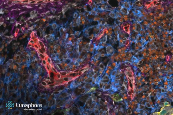
Another project focused on creating a multi-omic profile of high-grade serous ovarian cancer. Brodie integrated whole genome sequencing, multiplex IF, H&E staining, and transcriptomic data using the 10X Genomics Visium platform. Brodie mentioned that multiplex IF was conducted using the Ultivue workflow, which facilitated the analysis of 8 different protein markers in one image. Four clinically significant subtypes were found, and the tumour microenvironment for each one was analysed.
In conclusion, the successful implementation of these technologies has provided valuable insights into cancer biology. This work lays down important foundations in the spatial biology field, which could carve out a path for future research and potential therapeutic advancements.