Spatial cytometry refers to the tissue classification and assessment of spatial relationships, while cytometry leads to comprehensive phenotypical analysis at the single-cell resolution. Coralie Guerin, Head of Cytometry at Institut Curie, introduced spatial cytometry, a technology that combines tissue classification and single-cell resolution phenotypical analysis. A logarithmic view is needed to understand the universe and the human body comprehensively. It involves mapping the system at the single-cell resolution.
Guerin explained that the single-cell initiative at the Institut Curie unites four different platforms: the technology platform, the cytometry platform, the custom single-cell platform, and the bioinformatics platform to decipher spatial biology. This exciting initiative aims to implement solutions for simplex and multiplex protein measurement, targeted, and genome-wide RNA measurement, and multiomic analysis pipelines.
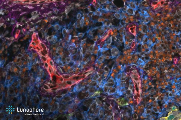
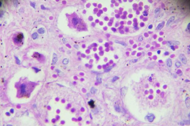
Guerin said the goal is to perform H&E imaging on the same slide used for multiplex staining. Rarecyte Orion is a tool dedicated to spatial cytometry. It was selected because it can acquire whole slides quickly with a single staining procedure. Furthermore, it is equipped with nine lasers and tunable filters allowing the measurement of up to 20 colours. The objective of the project was to apply the Orion to selected tissue samples.
The workflow involves staining slides with antibodies, obtaining the image, and processing data to obtain a single ome.TIFF file with corrected layers. Guerin explained that the stained slide is loaded, the acquisition with the software. She added that the software allows for customisation of the scan area, objective, channel, and colours. It is essential to conduct a few optimization steps such as antibody validation. The IHC method was picked for antibody validation because it doesn’t need any improvement regarding the fluorescence.
It is very important to acquire whole slides for studying tumour heterogeneity and other complex biological structures. Moreover, it allows researchers to explore how different regions of a tumour express various biomarkers.
Orion has been successfully applied to human and mouse tissue samples, including those from cancers such as head and neck, breast, and ovarian. Guerin also mentioned that its ability to subtract auto-fluorescence enhances the detection of weakly expressed proteins. So, the adaptability of the system allows researchers to design custom antibody panels tailored to specific projects.
The Orion spatial cytometry system provides a powerful, non-destructive, high-resolution approach to studying cellular interactions within tissues. This could have major implications for cancer research and pathology.