The Human Cell Atlas project is a consortium of groups collaborating to map all of the cell types in the human body, from in utero to aging. Cell types are defined by the genes they express which entails genomic and RNA sequencing work. At first, the atlas aims to investigate healthy human tissues and achieve a baseline understanding, before moving on to diseased tissues.
The consortium works across 95 countries with over 1500 institutes and 3000 members working towards mapping the cell types of the human body. Their mission is to enhance the understanding of the underlying biological mechanisms of disease, hopefully aiding drug discovery and the development of new medicines. Furthermore, the cell type atlas can provide an understanding of where a gene should be expressed and on which cell types. This gives drug developers an indication of where to look for possible side effects.
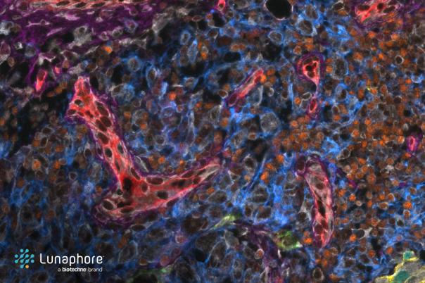
The Atlas employs two experimental branches: single cell and single nuclei sequencing and spatially resolved methods. Both of these branches are also followed by a heavy amount of bioinformatics. Wilbrey-Clark stressed the importance of keeping the data from their project open access for everyone in the scientific community to use.
Various spatial transcriptomics techniques are used, such as RNA scope, in situ sequencing, Xenium, Visium, and Curio. Each technique has its advantages and limitations, and the choice of method depends on the specific requirements of the study.
Wilbrey-Clark zoomed into the use cases and differences between Visium and Curio. Visium offers the advantage of H&E or immunofluorescence staining on the same section used for gene expression analysis and can work with low-quality samples using a probe-based method. However, it currently lacks single-cell resolution, which is expected to improve with future updates.
Curio, on the other hand, does not require tissue optimisation and is advertised as being more straightforward to use. However, it is not compatible with FFPE tissue and struggles with low-expressed genes. Additionally, the H&E staining is not performed on the same section as the gene expression analysis.